MRI KNEES SEGMENTATION
PROJECT COLLABORATORS
PUBLICATION
PROJECT BRIEF
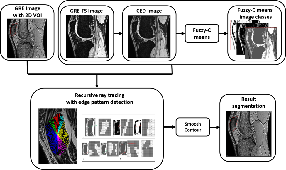
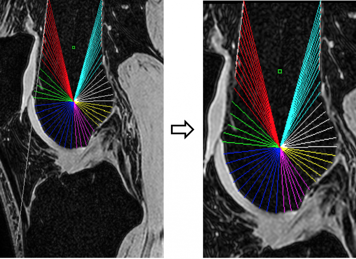
Through MIPAV plug-ins, BIRSS has collaborated with NIH IRP scientists on dozens of projects, helping to advance their work through agile development and a close working relationship. One such collaboration was the research BIRSS team members performed in concert with Dr. Frances Gavelli of National Institutes of Neurological Disorders and Stroke. BIRSS developers worked very closely with Dr. Gavelli and her team to design and implement as a MIPAV plug-in an automatic segmentation methodology for the patellar bone, based on 3D gradient recalled echo and gradient recalled echo with fat suppression magnetic resonance images. Constricted search space outlines are incorporated into recursive ray-tracing to segment the outer cortical bone. A statistical analysis based on the dependence of information in adjacent slices is used to limit the search in each image to between an outer and inner search region. A section based recursive ray-tracing mechanism is used to skip inner noise regions and detect the edge boundary. The proposed method achieves higher segmentation accuracy (0.23mm) than the other methods with the average dice similarity coefficient of 96.0% (SD 1.3%) agreement between the auto-segmentation and ground truth surfaces.
RESULTS
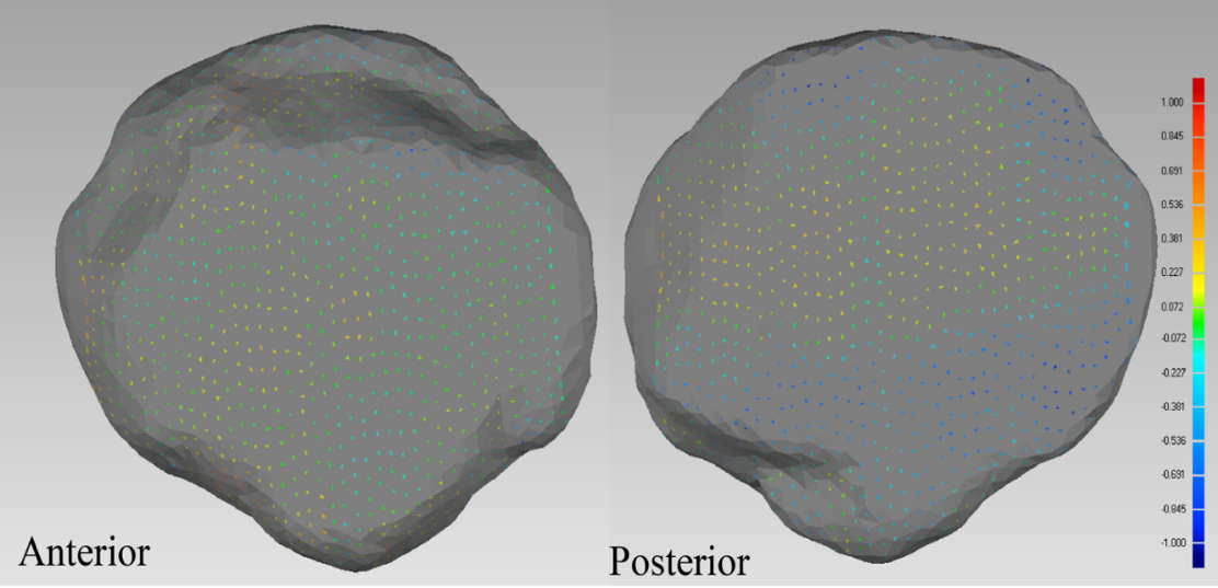
The average positive error was 0.25mm (SD 0.05mm)
The average negative error was -0.22mm (SD 0.17mm)
The average dice similarity coefficient was excellent, demonstrating 96.0% (SD 1.3%) agreement between the surfaces.
The average error was 0.02mm
The average positive error was 0.54mm
The average negative error was -0.46mm
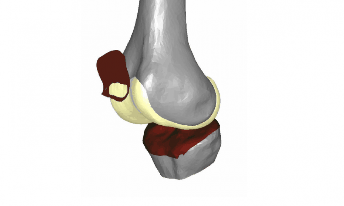
The bone and cartilage models were segmented in MIPAV and then using our registration process they can be animated to demonstrate cartilage contact patterns.
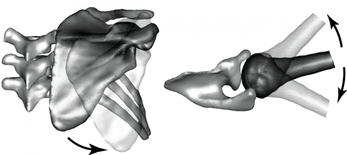
We are expanding this project to look at the shape of the scapula and to expand into motion.